It would be interesting to find parasitoids or predators to control Silba adipata McAlpine at certain stages of its life cycle. We report herafter the rare observations on this subject, but, as of the last modification date of this chapter (November 28, 2024), no species has been identified that could produce results large enough to halt the Silba adipata McAlpine.devastating action on immature figs.
F. SILVESTRI'S OBSERVATIONS
Filippo SILVESTRI, in its masterful study of the Black Fig Fly, indicates that he observed the presence in pupae of a tiny parasitoid hymenoptera (2 mm long), the name of which is Pachyneuron vindemmiae Rondani. Reference: SILVESTRI F., 1917, Sulla Lonchaea aristella Beck. (Diptera : Lonchaeidae) dannosa alle infiorescenze e fruttescenze del caprifico e del fico, Bollettino del Laboratorio di Zoologia Agraria in Portici, vol.12, pp. 123 -146.
The author specifies that the female of this species searches for the Black Fig Fly pupae which are on the surface of the soil between the dropped figs, or those wich are poorly hidden at a little depth, and deposits one egg on the nymph (not in the nymph) inside the pupa. The larva emerging from this egg feeds on the pupa of the Black Fig Fly, and, after having fully developed, transforms into a pupa inside the Black Fig Fly pupa. Then, the adult Pachyneuron vindemmiae Rondani emerges from its pupa and that of the Black Fig Fly by piercing a hole with
its mandibles. The complete development of the parasitoid, from egg to adult, takes 15 days in summer, and 16 to 23 days from September to November.
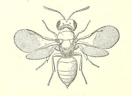
Pachyneuron vindemmiae Rondani (female specimen).
Credit: F. SILVESTRI - see description.
F. SILVESTRI considers that this species would be of negligible contribution in the Black Fig Fly control, because it cannot attack the almost totality of the pupae, wich are burried in the soil at 2-10 cm depth. Moreover, he mentions that he only found Pachyneuron vindemmiae Rondani in the Portici and Resina localities, whereas he examined thousands of pupae collected in the Napoli, Salerno and Lecce regions, and more than 1000 pupae received from Tripoli.
F. SILVESTRI does not know of any other specific enemy to the Black Fig Fly, including any bacteria or fungi. He simply notes that the blastophage can be considered at the level of the caprifig tree as an indirect enemy of the Black Fig Fly. Indeed, the blastophage is at the origin of the transformation of the flowers into galls which completely fill the central cavity of the fig, which prevents the young larvae which are there from moving and causes their death.
B. I. KATSOYANNOS' OBSERVATIONS
We found also a more recent reference to this parasitoid hymenoptera. B. I . KATSOYANNOS has studied large populations of black fig flies in the Chios Island (Greece), in 1981 and 1982. Reference: KATSOYANNOS B. I., 1983, Field observations on the biology and behavior of the black fig fly Silba adipata McAlpine (Diptera, Lonchaeidae), and trapping experiments, Z. ang. Entomol. 95, pp. 471-476. The author reports that he found (once only - September 27, 1981), among 70 collected pupae, 8 pupae of which emerged 8 specimens of the parasitoid species Pachycrepoideus vindemmiae Rondani (synonym of Pachyneuron vindemmiae Rondani).
E. TUTMUS' OBSERVATIONS
In his thesis relating to Silba adipata McAlpine, defended in 2013 at the University of Aydin (Turkey), Esref TUTMUS provides on page 32, § 4.5, some indications relating to the enemies of Silba adipata McAlpine. In 2011, no natural enemies of Silba adipata McAlpine were detected in figs. In 2012, parasitoid wasps and predatory ants were found. But the author does not provide any details, simply mentioning that an identification phase is underway. Reference: TUTMUS E., 2013, University Adnan MENDERES thesis - Aydin (Turkey), Aydin ili incir bahçelerinde Silba adipata McAlpine (=Lonchaea aristella Becker) (Diptera: Lonchaeidae)'nin yayilisi, popülasyon degisimi ve zarar oraninin belirlenmesi, 44 p.
J.H. GILIOMEE'S INDICATION
J.H. GILIOMEE et al. indicate that in South Africa, where Silba adipata McAlpine was identified in 2006 and 2007, no parasitoids were observed for the species. Reference: GILIOMEE J. H., E. VENTER E., WOHLFARTER M., 2007, Mediterranean black fig fly, Silba adipata McAlpine (Diptera: Lonchaeidae), recorded from South Africa, African Entomology 15(2), pp. 383-384.
ANTS: OURS OBSERVATIONS
F. DROUET has observed that the ants attack the eggs of Silba adipata McAlpine. They extract the eggs from the ostiole of immature figs, after having shredded the ostiolar scales, most often in small groups.
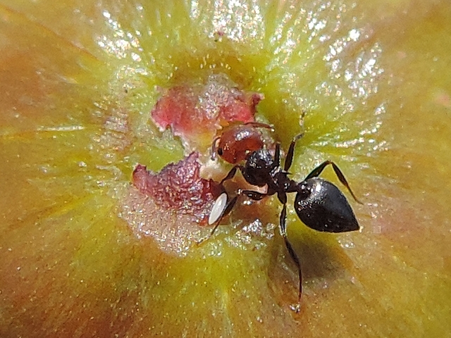
Ant having extracted a Black Fig Fly egg from the ostiolar scales.
Credit: F. DROUET.
Margaux ALLIX (Perpignan, France) and Alain COSTA (Albatera, Spain), two of the agonomists belonging to our small team, have observed that the ants attack the larvae of Silba adipata McAlpine.(when they have fallen to the ground, or when they are abandoning the immature fig).
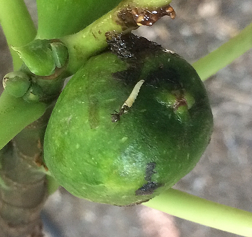
Ant attacking a Black Fig Fly larva, which is abandoning the immature fig.
Credt: Alain COSTA.
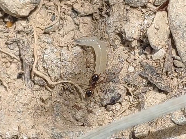
Ant attacking a Black Fig Fly larva, which fell fom the immature fig to the ground.
Credit: Margaux ALLIX (CIVAMBIO 66).
Should we consider ants as regulators of the Black Fig Fly populations ? Certainly, but unfortunately in a very limited way because, according to our observations, the percentages of Silba adipata McAlpine eggs and larvae removed by ants are very low.
B. PEYRE'S CURIOUS OBSERVATION
Communication from Bernard PEYRE, former fig producer, consultant-trainer for commercial fig cultivation.
On October 10, 2024, I collected seven ripe figs of the biferous variety 'Dalmatia' (greenish epidermis when ripe) from an orchard infested with Silba adipata McAlpine. They shew no larvae exit holes. In the central cavity of all these figs, I found mold on larvae damage, as well as pupae or dried blackish imagos of Silba adipata McAlpine. I collected from their reddish infructescence, for examination purposes, all the elements that could result from the Silba adipata McApine infestation. For one of these figs (numbered 4, photographs below), among the extracted elements was what appeared to me to be a very small whitish dead larva, which gave rise to a curious observation.
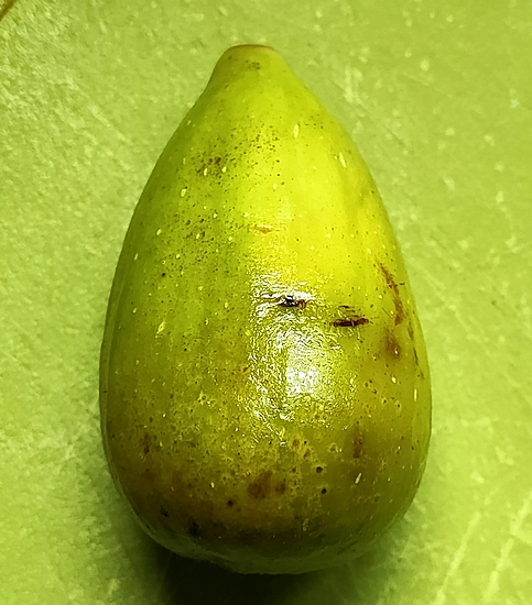
Whole 'Dalmatia' ripe fig no. 4.
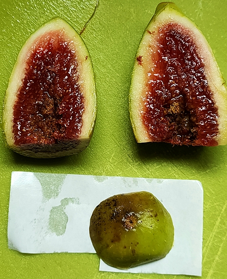
Interior of 'Dalmatia' ripe fig no. 4.
(the summit region was the subject of a transverse slice to search for chorions at the level of the ostiole.
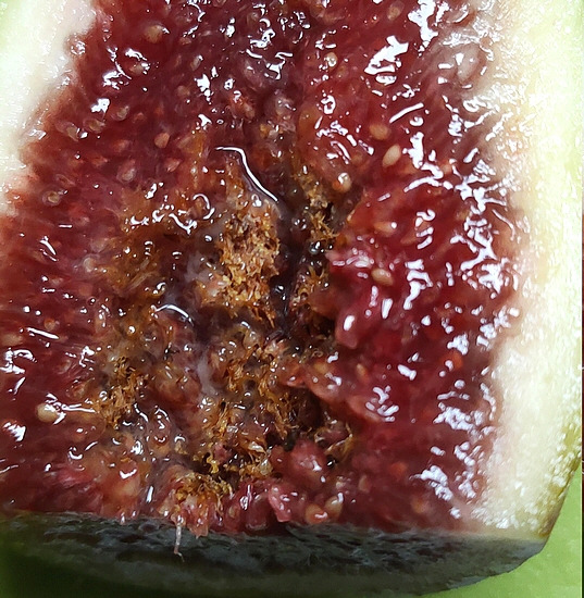
Mold on larvae damage within the infructescence of 'Dalmatia' ripe fig no. 4.

Elements extracted from the infructescence of 'Dalmatia' ripe fig no. 4.
(note the whitish element, which looks like a very small dead larva).
Examination of the ostiolar region of 'Dalmatia' ripe fig no. 4 under a stereomicroscope confirmed its infestation by Silba adipata McAlpine, revealing 12 chorions of this species, distributed under 5 ostiolar scales (2 + 5 + 3 + 1 + 1). It also revealed the presence of Drosophila suzukii Matsumura chorions (2 respiratory filaments) under the ostiolar scales and in the ostiolar canal, in small numbers. See examples in the photographs below.
Three Silba adipata McAlpine chorions found under an ostiolar scale of 'Dalmatia' ripe fig no. 4.
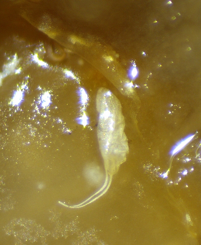
A Drosophila suzukii Matsumura chorion found under an ostiolar scale of 'Dalmatia' ripe fig no. 4.
(note: in actual size, this chorion is half the size of that of Silba adipata McAlpine).
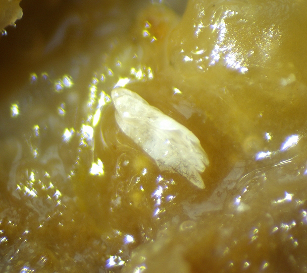
Concerning the whitish dried element extracted from the infructescence which suggested a very small larva, I was intrigued by its too compact shape (width too large in relation to length), as it appeared in my photographs taken on the table after removal from the infructescence. Examination under a stereomicroscope gives the answer for this unusual shape, but reveals a very rare observation in the form of an enigma (see photograph below).
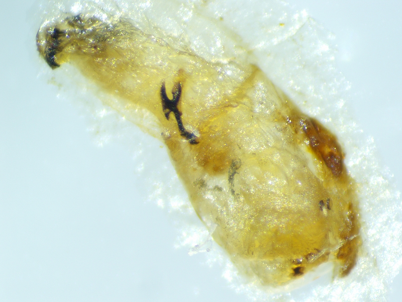
Examination under a stereomicroscope of the whitish dried element extracted from the infructescence of 'Dalmatia' ripe fig no. 4.
I received the contribution of François DROUET for the interpretation of the photograph. For us, it is in fact a larva (the anterior end and part of the body of which are visible) which seems to have died before managing to completely extract itself from a much larger whitish host larva (of which only the posterior part remains, on the right in the photograph). Given the state of drying of the examined element, it was not possible to identify posterior spiracles. The clue which supports the fact that these are two larvae is the presence of a second pharyngeal armature, completed with mandibles, along the body of the emerging larva. It should be noted that this pharyngeal armature is about twice the size of the pharyngeal armature of the emerging larva. Based on the size of the overall examined element (2 mm; photograph below), the emerging larva could measure between 1.5 and 2 mm. The attempt to extract the larva failed, because this one broke due to drying out. The size of the host larva is more difficult to assess from the remaining fragment (3 mm?).
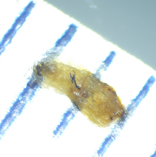
Size of the whitish element extracted from the infructescence of 'Dalmatia' ripe fig no. 4.
(measured size: 2 mm).
Our hypothesis is that it is a larva of a parasitoid that has not managed to completely extract itself from the remains of a host larva. Without knowing which two species are involved. We also do not know wether the parasitoid larva only emerges once fully developed. If it does, the corresponding imago is a very small insect. And we do not know at what age the host larva was attacked. If the host larva measured 3 mm at the time of the attack, and the attack blocked its development, it could be a 2-day-old Silba adipata McAlpine larva. If the development of the host larva was blocked only after incubation of the parasitoid egg (1 day?), the host larva may have been 2 mm in size at the time of attack, which would correspond to a 1-day-old Silba adipata McAlpine larva. But the host larva may also belong to a species other than Silba adipata McAlpine.
 Top of page Summary