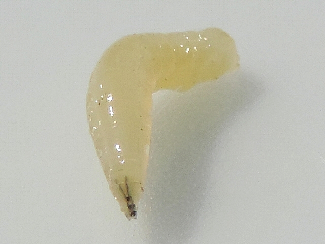
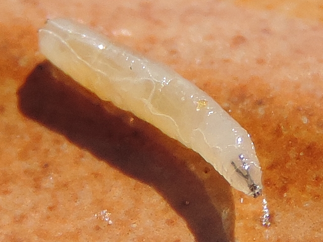

RESPIRATORY SYSTEM
SPIRACLES
The mature larva has two pairs of spiracles (respiratory orifices): an anterior pair, and a posterior pair (not visible to the naked eye). The newly born larva has only two spiracles (the posterior ones).The anterior spiracles are located on the first thoracic segment
(in its posterior part, at the limit with the second segment). The posterior spiracles are located on the upper part of the larva rear, and are much larger than the anterior ones.
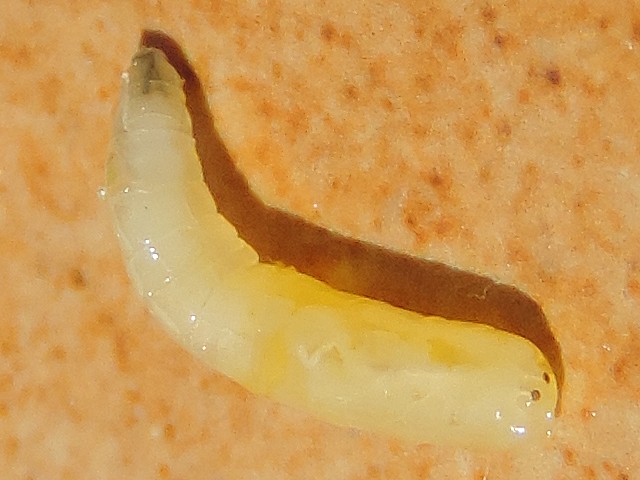
Silba adipata McAlpine larva: the four spiracles.
POSTERIOR SPIRACLES
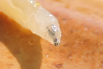
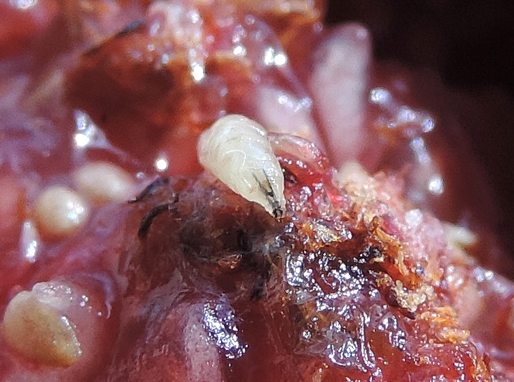
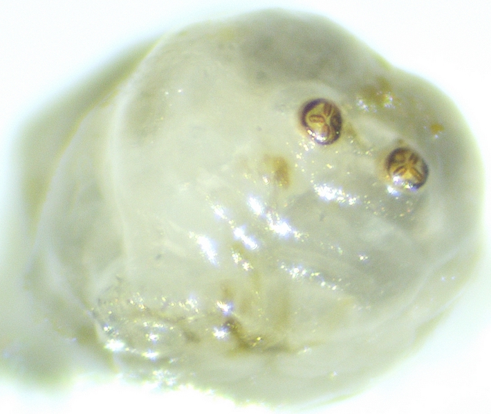
At the larva posterior end, we observe two brown spiracles in a dorsal position on the truncated part of the last segment.

Silba adipata McAlpine larva: posterior spiracles.
(larvae in a fig central cavity, among the devoured flowers ends wich have turned brown).
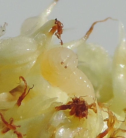
Silba adipata McAlpine larva: posterior spiracles.
(larva photographed in the partly devoured and browned inflorescence of the fig).
On page 129 of his masterful study relative to the Black Fig Fly, F. SILVESTRI gives some details. The posterior spiracle is carried by a very short cylindrical protuberance, and shows three lateral slits. Each of the slits is elongated and provided on its inner limit with small pea-shaped protuberances forming a filter for the air. On the spiracle plate, there are four branched bristle-like appendages. And the author provides the drawing below. Reference: SILVESTRI F., 1917, Sulla Lonchaea aristella Beck. (Diptera : Lonchaeidae) dannosa alle infiorescenze e fruttescenze del caprifico e del fico, Bollettino del Laboratorio di Zoologia Agraria in Portici, vol.12, pp. 123 -146.
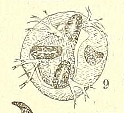
Silba adipata McAlpine larva: posterior spiracle details.
Credit: F. SILVESTRI.
JUÁREZ MAYA M. et al. have published in the Biodiversity Data Journal of November 2024 an annotated photograph of the Silba adipata McAlpine larva posterior spiracle, which confirms in every respect the drawing and the description of F. SILVESTRI. Reference: JUÁREZ MAYA M., MORALES GALVÁN Ó., VALDEZ CARRASCO J., ILLESCAS SALVADOR D., GUTIERREZ RUELAS J., ILLESCAS RIQUELME C., 2024, Morphology of immature stages of the black fig fly Silba adipata (Diptera, Lonchaeidae), Biodiversity Data Journal 12: e137798.
Photograph shown below (authorized reproduction, CC BY 4.0 license), in which: SS = spiracular slit; SSC = stigmatic scar, without orifice (also called the button); ISP = interspiracular processes (four in number, I to IV). Note that the location and orientation of the four hyaline processes are strictly the same in the photograph and in the drawing by F. SILVESTRI.
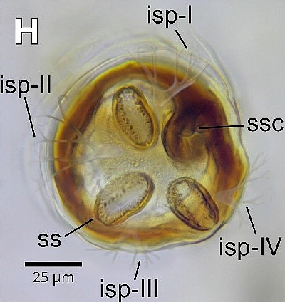
Silba adipata McAlpine larva: posterior spiracle details.
Credit: JUÁREZ MAYA M. et al. (Biodiversity Data Journal).
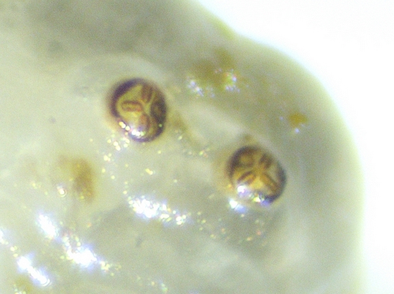
When I observe the larva posterior spiracles under the stereomicroscope, only the respiratory slits configuration is visible. It should be noted that two of them are in the extension of each other, the third being perpendicular to the other two. Opposite the third slit, a more or less circular spot appears, light
to quite dark depending on the light incidence (photograph below). Let us emphasize that this posterior spiracles configuration is the identification criterion that I have retained to distinguish in ripe figs the larvae of Silba adipata McAlpine from those of Ceratitis capitata Wiedemann (see the chapter
on the larvae species identification in ripe figs).
Silba adipata McAlpine larva: posterior spiracles.
Silba adipata McAlpine larva: posterior spiracles.
ANTERIOR SPIRACLES
The larva anterior spiracles are located in an upper lateral position, in the rear area of the first thoracic segment, almost at the limit with the second thoracic segment. I was able (with difficulty) to observe them in only a few photographs.
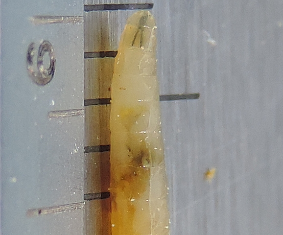
Silba adipata McAlpine larva: anterior spiracle on the right side, at the level of the first thoracic segment.
(it is the tiny white protuberance almost at the limit with the second segment, from where the right main trachea starts).
On page 129 of his masterful study relative to the Black Fig Fly, F. SILVESTRI gives some details. The anterior spiracle protudes little from the surface of the larva body, and has 8 or 9 lobes, sometimes 7. And the author provides the drawing below. Reference: SILVESTRI
F., 1917, Sulla Lonchaea aristella Beck. (Diptera : Lonchaeidae) dannosa alle infiorescenze e fruttescenze del caprifico
e del
fico, Bollettino del Laboratorio di Zoologia Agraria in Portici, vol.12, pp. 123 -146.
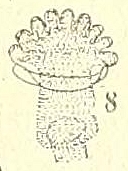
Silba adipata McAlpine larva: anterior spiracle details.
(note the lobes through which air enters the main trachea).
Credit: F. SILVESTRI.
JUÁREZ MAYA M. et al. have published in the Biodiversity Data Journal of November 2024 a photograph of the Silba adipata McAlpine larva anterior spiracle, which confirms the description of F. SILVESTRI. Reference: JUÁREZ MAYA M., MORALES GALVÁN Ó., VALDEZ CARRASCO J., ILLESCAS SALVADOR D., GUTIERREZ RUELAS J., ILLESCAS RIQUELME C., 2024, Morphology of immature stages of the black fig fly Silba adipata (Diptera, Lonchaeidae), Biodiversity Data Journal 12: e137798. Photograph shown below (authorized reproduction, CC BY 4.0 license), in which the anterior spiracle has 9 lobes.
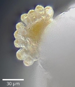
Silba adipata McAlpine larva: anterior spiracle.
Credit: JUÁREZ MAYA M. et al. (Biodiversity Data Journal).
When I observe the larva body under the stereomicroscope, I easily spot the anterior spiracle, but its details are not really visible.
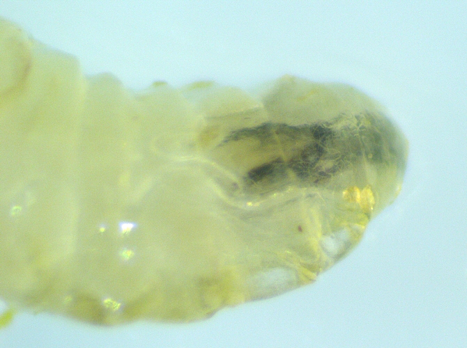
Silba adipata McAlpine larva: right anterior spiracle.
(location: posterior part of the first segment, at the limit with the second segment).

Silba adipata McAlpine larva: anterior spiracles.
(note: lobes are visible for the right spiracle; the left spiracle appears as a small black bar).
TRACHEAS
Two dorsal longitudinal trunks (main tracheas) extend between the anterior and posterior spiracles (one on each side). From the main tracheas, a complex tracheal branches network supplies air to the muscles and the internal organs. In the photograh below, we can follow the left main trachea from the left anterior spiracle to the left posterior one.
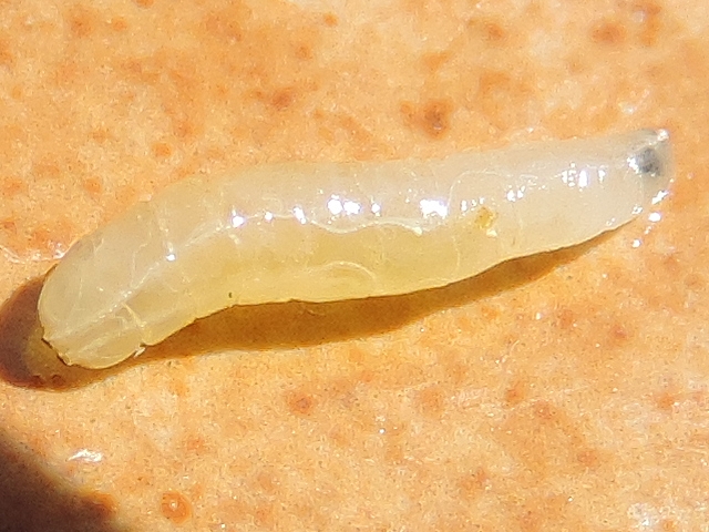
Silba adipata McAlpine larva: main tracheas.
In this second photograph, we can see the exact location of the left anterior spiracle on the first thoracic segment. And we can follow the main trachea starting from it, to join the posterior spiracle (not visible).
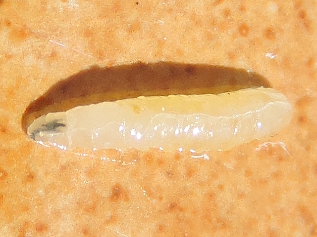
Silba adipata McAlpine larva: left main trachea and left anterior spiracle.
This third photograph shows the junction of the right main trachea with the corresponding anterior spiracle.

Silba adipata McAlpine larva: junction of the right main trachea with the corresponding anterior spiracle.
And the photograph below shows some ramifications of the right main trachea.
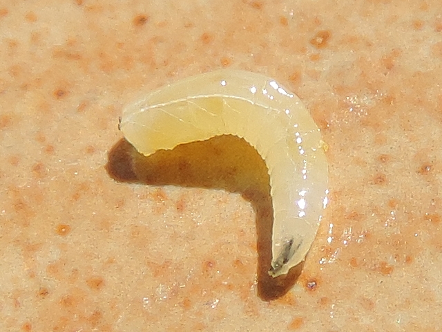
Silba adipata McAlpine larva: sone ramifications of the right main trachea.
VENTRAL SURFACE
CREEPING WELTS
The larva's ventral surface is characterized by the presence of creeping welts (fleshy transverse ridges), which assist it in its movements.

Silba adipata McAlpine larva: creeping welts (the last relef, on the left, is the anal area).
Below, a photograph showing the ventral surface of a Silba adipata McAlpine larva. From left to right in the photograph, three features can be highlighted, as follows. The ventral surface of the three first segments (thorax) is smooth, they have no creeping welts. The creeping welts of the eight abdominal segments are located on their anterior part, and are
increasingly big from the first to the last of them. The last segment shows two protuberances: a creeping welt in its anterior part, and the anal area in its posterior part.

Silba adipata McAlpine larva: ventral surface.
On page 129 of his masterful study relative to the Black Fig Fly, F. SILVESTRI gives some details about the creeping welts. Each of the eight abdominal segments has a transverse elevation, located in its anterior part. The elevation of the first abdominal segment has 3 to 4 transverse rows of points of equal height; the elevation of the other abdominal segments has 3 transverse rows of points, the latter higher in the middle row. Reference: SILVESTRI F., 1917, Sulla Lonchaea aristella Beck. (Diptera : Lonchaeidae) dannosa alle infiorescenze e fruttescenze del caprifico e del fico, Bollettino del Laboratorio di Zoologia Agraria in Portici, vol.12, pp. 123 -146.
ANAL AREA
The anal area is circular
and has two bifid lobes.
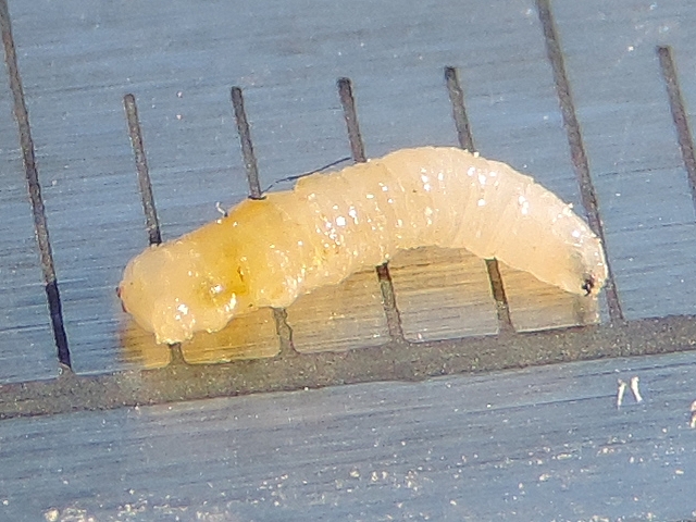
Silba adipata McAlpine larva: anal area (circular relief just on the second millimeter graduation, on the left).
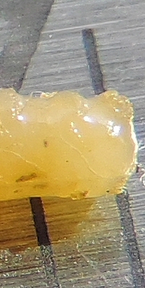
Silba adipata McAlpine larva: anal area, circular, with two bifid lobes.
(preceded by the creeping welt of the last segment, and that of the penultimate one).
On page 129 of his masterful study relative to the Black Fig Fly, F. SILVESTRI specifies that there is a small circular area surrounding the anal valves, which is provided with strong spinules in 2-3 irregular rows behind the anus, and in 2 rows in front of it. Reference: SILVESTRI F., 1917, Sulla Lonchaea aristella Beck. (Diptera : Lonchaeidae) dannosa alle infiorescenze e fruttescenze del caprifico e del fico, Bollettino del Laboratorio di Zoologia Agraria in Portici, vol.12, pp. 123 -146.
JUÁREZ MAYA M. et al. have published in the Biodiversity Data Journal of November 2024 a photograph of the Silba adipata McAlpine larva anal area. Reference: JUÁREZ MAYA M., MORALES GALVÁN Ó., VALDEZ CARRASCO J., ILLESCAS SALVADOR D., GUTIERREZ RUELAS J., ILLESCAS RIQUELME C., 2024, Morphology of immature stages of the black fig fly Silba adipata (Diptera, Lonchaeidae), Biodiversity Data Journal 12: e137798. Photograph shown below (authorized reproduction, CC BY 4.0 license), in which we can note that the two lobes are bifid (with a rear part smaller than the front one), and we can distinguish the posterior irregular rows of thick sclerotized spinules.
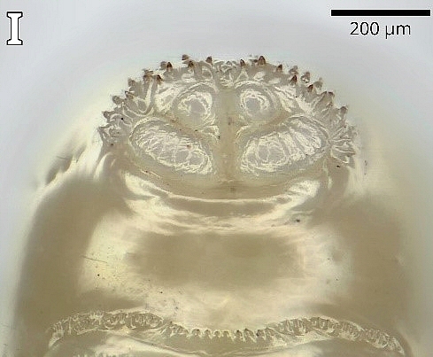
Silba adipata McAlpine larva: anal area, on the ventral surface (turned over).
(at the bottom, the three rows of points of the last segment creeping welt).
Credit: JUÁREZ MAYA M. et al. (Biodiversity Data Journal).
