



|
Presentation Biology Living habits Infestation Control methods |
||
|
|
||
|
|
|
Author: François DROUET. |
|
|
||
|
The larva: morphology, size, cephalic capsule
I give the Black Fig Fly larva description, according to the following plan: part 1 (this chapter): morphology, size, cephalic capsule; part 2 (see chapter): pharyngeal armature, respiratory system, ventral surface.
MORPHOLOGY
The larva is formed of 11 segments (3 for the thorax, 8 for the abdomen), plus a cephalic capsule (head) which
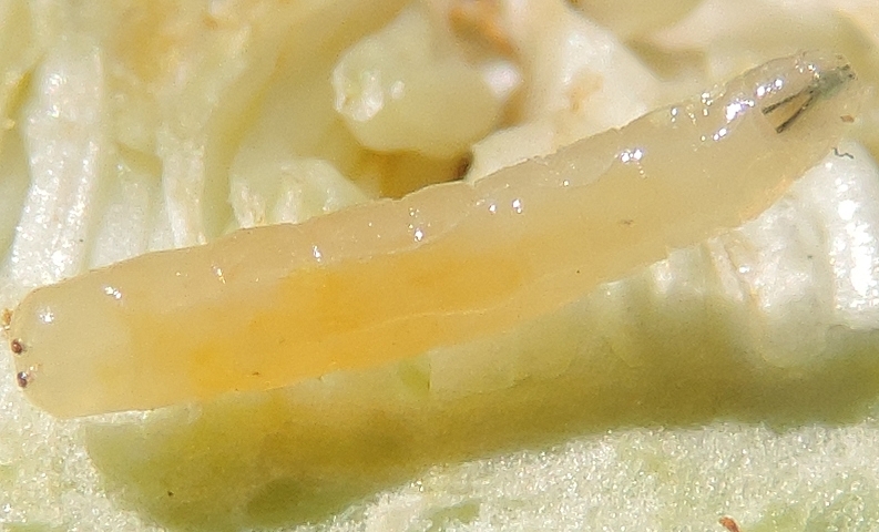
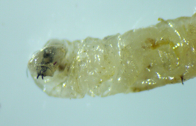
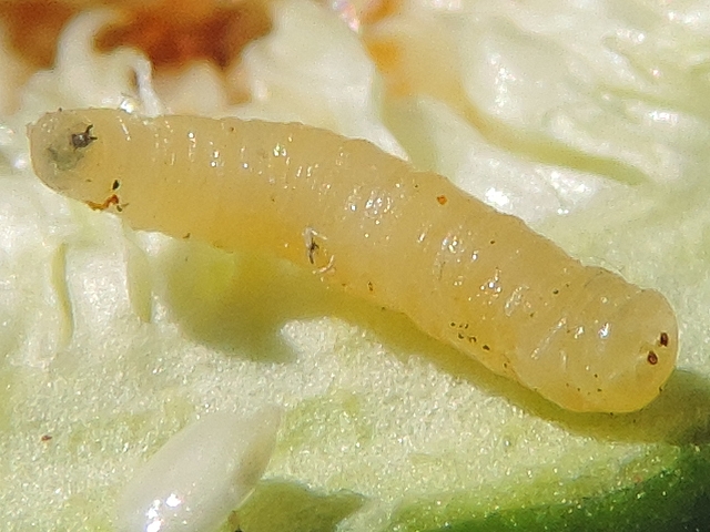
is retractable inside the first thoracic segment. In the past, the anterior end of the flies larva was called the "head", but contemporary entomologists classify the flies larva among the acephalic larvae and identify a "cephalic capsule". In the photograph 1, below, the cephalic capsule is not visible, because
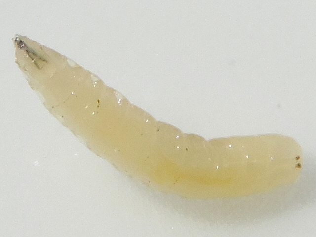
its is retracted inside the first thoracic segment. The latter is easily recognizable having a different pointed shape compared to the other segments - on the right in the photograph.
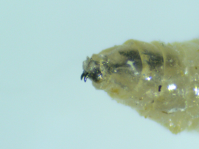
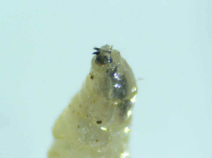
Black Fig Fly larva with the cephalic capsule retracted inside the first thoracic segment. In the photograph 2, below, the tiny cephalic capsule is visible, protuding from the first thoracic segment - on the right in the photograph. It is not visible to the naked eye.
Black Fig Fly larva with the tiny cephalic capsule protubing from the first thoracic segment. On the last segment of the larva, we can see two brown dots, which are respiratory orifices called the posterior spiracles - on the left in the two photographs above. They are not visible to the naked eye. Inside the two first thoracic segments, we observe by transparency a black Y-shaped anatomical piece composed of the pharyngeal armature extended by the mandibles (mouth hooks). The pharyngeal armature is a skeleton on which certain muscles are attached, in particular those of the pump which sucks food.
SIZE
When I examine the ostiolar scales of immature figs with the stereomicroscope, it is not uncommon that I find Silba adipata McAlpine larvae which have just emerged from the eggs, and which have not yet begun their journey towards the fig central cavity. According to my measurements, the length at birth of the Silba adipata McAlpine larva is a little less than 1 mm. F. SILVESTRI indicates more precisely a length of 0.80 mm and a width of 0.18 mm. Reference: SILVESTRI F., 1917, Sulla Lonchaea aristella Beck. (Diptera : Lonchaeidae) dannosa alle infiorescenze e fruttescenze del0 caprifico
e del
fico, Bollettino del Laboratorio di Zoologia Agraria in Portici, vol.12, pp. 123 -146.
Silba adipata McAlpine larva under an immature fig ostiolar scale.
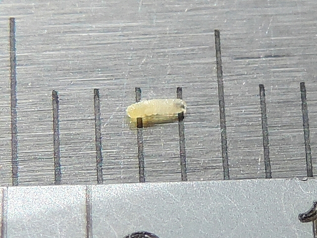
Silba adipata McAlpine larva size at birth.
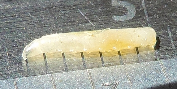
Silba adipata McAlpine larva size at birth. And the larva length is about 7mm at full development (when it leaves the unripe fig). Knowing that when we cut and open an old infested fig, the largest larvae are most frequently 6.5 mm long, not having quite completed their development.
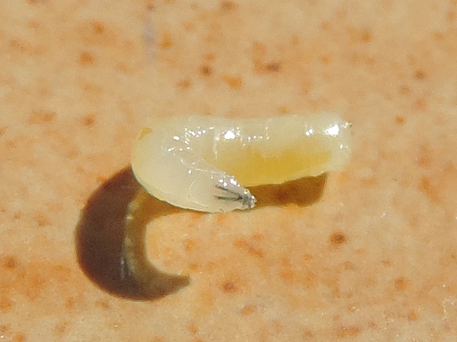
Silba adipata McAlpine larva maximum length (7 mm). Regarding the smallest larvae that I observed in the fig central cavity, the minimum length that I measured is 1.5 mm. The smaller larvae (1 mm long) having escaped my observation during my researches in the central cavity of the figs, yet numerous. The discovery in the fig central cavity of a 1.5 mm long larva is an extremely rare observation. When an immature fig is opened or fragmented to look for larvae, those of very small size (1-2 mm) remain inside the half-fig or fragments, flattened against the flowers, almost motionless due to their very low vivacity.
I noted 1.5 mm long larvae observations on April 16, 2020 and May 7, 2021, in breba figs of the 'Grise de la Saint-Jean' variety. The second observation was made possible only because the 1.5 mm long larva was stuck to a 4 mm long larva, when the latter fell on the observation dish, during the fig fragmentation with the fingers. At these times of the year, the size of 1.5 mm corresponds to a 2-day-old larva.
Silba adipata McAlpine larva 1.5 mm long (vey rare observation).
CEPHALIC CAPSULE
RETRACTILITY The cephalic capsule is retractile, and is often fully inserted into the body. More précisely in the first thoracic segment, which has a pointed shape, unlike the other ten larva segments. When the cephalic capsule is retracted, the first thoracic segment can be misinterpreted as what would constitute a head for the larva due to its pointed shape. But it is not so, we only see the thorax and the abdomen of the larva.
Black Fig Fly larva: cephalic capsule protuding from the first thoracic segment. The cephalic capsule is housed at the tip of the first thoracic segment in a sort of open sac (pocket), and projects forward when released.
Black Fig Fly larva: opening of the first thoracic segment pocket housing the cephalic capsule.
Black Fig Fly larva: cephalic capsule projected out of the first thoracic segment.
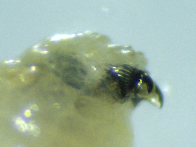
SIZE The cephalic capsule is of tiny size compared to that of the constituent segments of the larva, in particular of the first thoracic segment which shelters it. For the untrained eye, there is a risk of confusion with the terminal part of the first thoracic segment. We can notice that the first thoracic segment is in fact made up of two parts: a first part, pointed but wide, which follows the second thoracic segment; a second part, very short and much narrower after a clearly identifiable tightening, at the end of which appears the cephalic capsule when it is not retracted. The narrowed terminal part of the first thoracic segment should not be confused with the cephalic capsule. So, in the photograph below, the cephalic capsule is the tiny rounded appendage that appears at the very end of the larva anterior part. The pharyngeal armature rear points protrude into the second thoracic segment. If we move towards the larva rear from the cephalic capsule, we distinguish the terminal part of the first thoracic segment, very short and narrowed, which stops at the level where the two pharyngeal armature rear points (forming a V) meet; then the first part of the first thoracic segment, in the shape of a point but clearly wider.
Silba adipata McAlpine larva: tiny cephalic capsule protuding from the first thoracic segment.
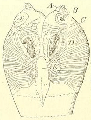
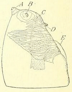
DESCRIPTION OF F.SILVESTRI F. SILVESTRI provides the two figures below for the Black Fig Fy cephalic capsule (which he calls the head). He highlights a bilobed form at the top, two antennae, a mouth, and two mandibles (mouth hooks). Reference: SILVESTRI F., 1917, Sulla Lonchaea aristella Beck. (Diptera : Lonchaeidae) dannosa alle infiorescenze e fruttescenze del caprifico
e del
fico, Bollettino del Laboratorio di Zoologia Agraria in Portici, vol.12, pp. 123 -146.
Black Fig Fly larva: cephalic capsule (viewed from below, left; wiewed laterally, right).
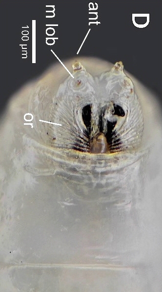
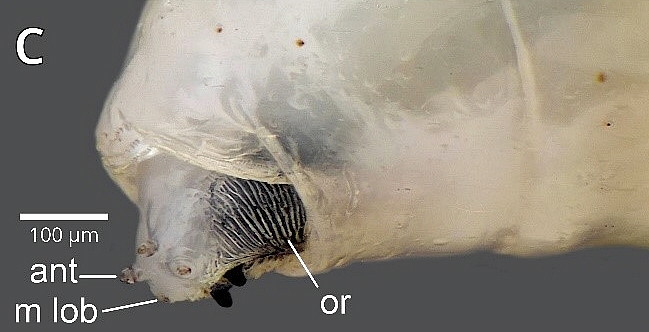
PHOTOGRAPHS OF JUÁREZ MAYA M. ET AL. Photographs provided by JUÁREZ MAYA M. et al. in the November 2024 Biodiversity Data Journal allow us to apprehend some details of the Silba adipata McAlpine larva cephalic capsule. Reference: JUÁREZ MAYA M., MORALES GALVÁN Ó., VALDEZ CARRASCO J., ILLESCAS SALVADOR D., GUTIERREZ RUELAS J., ILLESCAS RIQUELME C., 2024, Morphology of immature stages of the black fig fly Silba adipata (Diptera, Lonchaeidae), Biodiversity Data Journal 12: e137798. The two photographs below, taken from the aforementioned article (authorized reproduction, CC BY 4.0 license), show the cephalic capsule of the Silba adipata McAlpine larva in view from below (D) and lateral (C). The authors call the maxillary lobe, without specifying its function, the element named the antenna ventral part by F. SILVESTRI. Note: on D, the lower lip below the mouth hooks; on D (at the bottom) and on C (on the right), the groove delimiting the first thoracic segment in which the cephalic capsule is housed.
Black Fig Fly larva: cephalic capsule protuding from the first thoracic segment.
Black Fig Fly larva: cephalic capsule protuding from the first thoracic segment.
MOUTH F. SILVESTRI indicates that the mouth is located on the ventral part of the retractile cephalic capsule. The mouth orifice is not visible to the naked eye.
The author specifies that the oral lobes are located on the ventral surface and partly on the side, and that they show dense transverse lines (see the figures provided above). I was able to observe the oral lobes lines under a stereomicroscope (see photographs below).
Black Fig Fly larva: oral lobe with transverse lines.
Black Fig Fly larva: oral lobe with transverse lines.
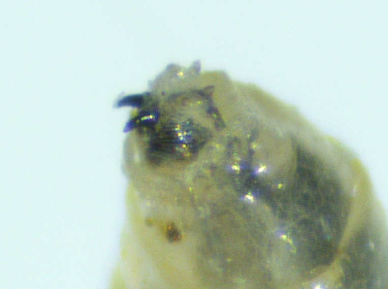
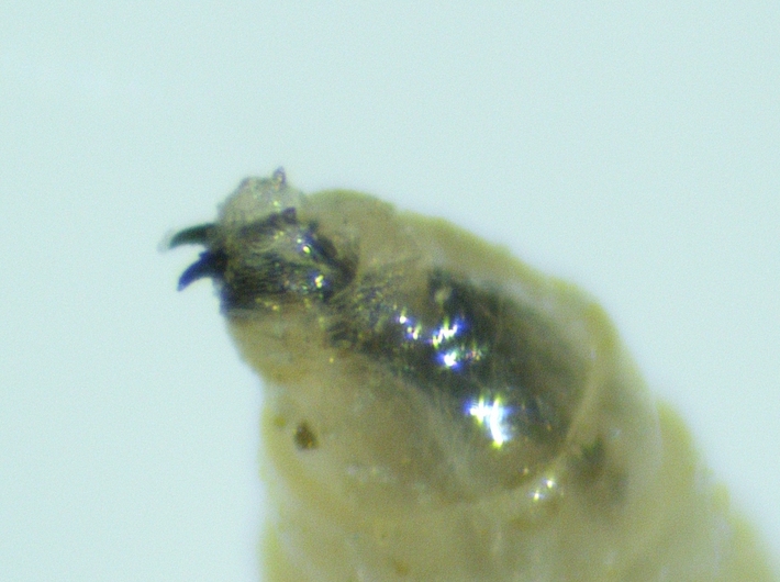
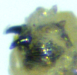
MOUTH HOOKS Above the lower lip are two mouth hooks. These play in the vertical plane and, according to certain entomologists, would also serve as locomotion organs. On the side of the inward part of the mouth hooks, the oral lobes end in a slightly lobed blade, also represented by F. SILVESTRI in the figures above. Below, photographs showing the two mouth hooks.. Black Fig Fly larva mouth hooks protubing from the first thoracic segment.
Black Fig Fly larva mouth hooks protubing from the first thoracic segment. Looking closely at the photograph below, we can see the lower lip, between the two mouth hooks, below the exit points of these.
Black Fig Fly larva: mouth hooks and lower lip.
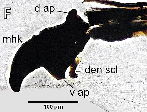
DETAIL OF THE MOUTH HOOKS The mouth hooks are the mandibles, the greater part of which is not externalized. The term mouthl hook refers to the entire mandible, but in descriptions, figures, or photographs, where only the external curved tip of the mandible appears, the term mouth hook refers to it. The mandible is a highly sclerotized anatomical piece, which has a rectangular internal basal part, with a rounded dorsal apodeme and a ventral apodeme having a pointed end directed backwards. It extends to the front by a narrower, curved, smooth part, ending in a rounded point. The mandible has a notch on its upper margin, between the dorsal apodeme and the end of the point. A curved dental sclerite is located behind the ventral apodeme. Mandibles are attached to the pharingeal armature by an intermediate sclerite (for the pharyngeal armature, see details in the second chapter devoted to the larva description).
See photograph below, taken from an article of JUÁREZ MAYA M. et al. published in the Biodiversity Data Journal of November 2024. Reference: JUÁREZ MAYA M., MORALES GALVÁN Ó., VALDEZ CARRASCO J., ILLESCAS SALVADOR D., GUTIERREZ RUELAS J., ILLESCAS RIQUELME C., 2024, Morphology of immature stages of the black fig fly Silba adipata (Diptera, Lonchaeidae), Biodiversity Data Journal 12: e137798. Authorized reproduction, CC BY 4.0 license.
Black Fig Fly larva: mandible, attached on the right to the intermediate sclerite.
ANTENNAE F. SILVESTRI indicates that the cephalic capsule bears two antennae, located above the mouth hooks, each at the top of one of the two lobes.
Black Fig Fly larva: two antennae, each of them at the top of one cephalic capsule lobe. The author specifies that each antenna consists of a point (composed of two very short articles) in its dorsal part, and of a slight protuberance in a circle in its ventral part (see figures provided above, and photograph below).
Black Fig Fly larva: details of cephalic capsule antennae.
Black Fig Fly larva: the antennae, two tiny points at the anrterior extremity.
|
||
